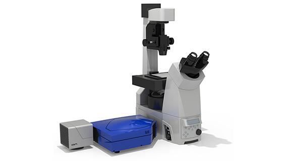
Confocal and spinning disk microscopy For live cell imaging
Scanning spinning disk confocal microscopy are exceedingly popular imaging techniques in cellular biology for fixed cell and in vivo imaging. In these modalities, using adaptive optics allows the user to increase the fluorescence signal and image contrast, especially when a high-magnification oil immersion objective is used to image water-based biological samples. Fixed biological samples, primarily imaged using scanning confocal microscopy, induce complex aberrations which distort the image and reduce resolution and sharpness.
Adaptive optics’ increasing of the signal-to-noise ratio can combat this distortion and reduction. When imaging deeper in biological samples with the spinning disk technique, correction of aberrations using adaptive optics more than doubles the fluorescence signal. This also allows the PSF to be uniform in different parts of the sample, which dramatically improves the quality of image deconvolution when post-processing the data.
This process allows the corrections for aberrations by gradually increasing with depth the amplitude of the correctionand It does not require illumination of the sample for aberration detection.


